Showing posts with label pharma-D-lab manual. Show all posts

Tuesday, December 22, 2009

Detailed study of frog- pharma-d labmanual

AIM:
To:
Describe the appearance of various organs found in the frog.
• Name locate and identify the organs that make up various systems of the frog.
• Compare and contrast frog anatomy to our past dissections.
• Contrast and compare various frog's organs to human.
Background information:
Frogs are classified as amphibians "live a double life". Frogs are part of the amphibian order, Anura. Tadpoles are aquatic and herbivores. Adult frogs can live on land and in water and are carnivores. Strong muscles and special fused bones help frogs be powerful swimmers and jumpers. Frogs have loose, mucous lined skin to help them escape from predators, and keep them wet which aides in cutaneous respiration (breathing through the skin). Tadpoles breath through gills. Frogs breath though underdeveloped lungs and their skin. Cutaneous respiration limits the frogs body size. The backs of frogs are dark, while their undersides are light, to camouflage them on land and water. Frog brains are smaller and less developed than other vertebrates, they also have a 3 chambered heart.

Materials required:
• safety goggles, gloves, and a lab apron
• forceps
• preserved frog
• dissecting pins (6–10)
• dissecting tray and paper towels
• plastic storage bag and twist tie
• scissors
• marking pen
• dissecting needle

Procedure:

1.Put on safety goggles, gloves, and a lab apron.
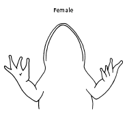
2.Place a frog on a dissection tray. To determine the frog’s sex, look at the hand digits, or fingers, on its forelegs. A male frog usually has thick pads on its "thumbs," which is one external difference between the sexes, as shown in the diagram below. Male frogs are also usually smaller than female frogs. Observe several frogs to see the difference between males and females.


3.Use the diagram below to locate and identify the external features of the head. Find the mouth, external nares, tympani, eyes, and nictitating membranes


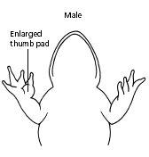
Turn the frog on its back and pin down the legs. Cut the hinges of the mouth and open it wide. Use the diagram below to locate and identify the structures inside the mouth. Use a probe to help find each part: the vomerine teeth, the maxillary teeth, the internal nares, the tongue, the openings to the Eustachian tubes, the esophagus, the pharynx, and the slit-like glottis.


5. Look for the opening to the frog’s cloaca, located between the hind legs. Use forceps to lift the skin and use scissors to cut along the center of the body from the cloaca to the lip. Turn back the skin, cut toward the side at each leg, and pin the skin flat. The diagram above shows how to make these cuts
6. Lift and cut through the muscles and breast bone to open up the body cavity. If your frog is a female, the abdominal cavity may be filled with dark-colored eggs. If so, remove the eggs on one side so you can see the organs underlying them.
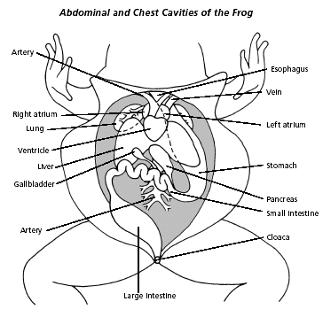
7.use the diagram below to locate and identify the organs of the digestive system: esophagus, stomach, small intestine, large intestine, cloaca, liver, gallbladder, and pancreas.
8.Again refer to the diagram below to identify the parts of the circulatory and respiratory systems that are in the chest cavity. Find the left atrium, right atrium, and ventricle of the heart. Find an artery attached to the heart and another artery near the backbone. Find a vein near one of the shoulders. Find the two lungs.
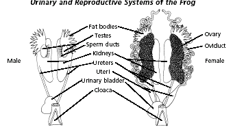
Use a probe and scissors to lift and remove the intestines and liver. Use the diagram on the next page to identify the parts of the urinary and reproductive systems. Remove the peritoneal membrane, which is connective tissue that lies on top of the red kidneys. Observe the yellow fat bodies that are attached to the kidneys. Find the ureters; the urinary bladder; the testes and sperm ducts in the male; and the ovaries, oviducts, and uteri in the female.


10.

Remove the kidneys and look for threadlike spinal nerves that extend from the spinal cord. Dissect a thigh, and trace one nerve into a leg muscle. Note the size and texture of the leg muscles.
11.

Dispose of your materials according to the directions from your teacher.
12.

Clean up your work area and wash your hands before leaving the lab.

Monday, December 21, 2009

STUDY OF TRANSVERSE SECTIONS-pharm-D labmanual

STUDY OF TRANSVERSE SECTIONS
AIM:
To study the transverse section of Senna, cassia, ephedra, Podophyllum
Principle:
A segment of the body that is at right angles to the body's longitudinal axis.
Senna the sennas, is a large genus of around 250 species of flowering plants in the family Fabaceae, subfamily Caesalpinioideae.
Almost all species were at one time or another placed in Cassia, a close relative which until recent decades served as a "wastebin taxon" to hold all Cassiinae
Ephedra is the dried herbaceous stem of Ephedra sinica Stapf, Ephedra intermedia Schrenk et C. A. Mey. or Ephedra equisetina Bge. (Fam. Ephedraceae)
Action: To induce perspiration for dispelling cold, to relieve asthma, and to cause diuresis
Podophyllum peltatum, Linne (N.O. Berberidece), is a small herb with a long perennial creeping rhizome.
The drug was well known to the North American Indians as an emetic and vermifuge;

Glassware requirement:
a small beaker
forceps
biological leaf cutter/blade
microscopic slide
distilled water

PROCEDURE:
Take the leaf of senna or cassia
wash it carefully with distilled water
take out from the beaker using forceps
using a sterile blade slice out the leaf carefully
obtain thin slice of leaf
mount in on to the microscopic slide carefully
observe it under microscope clearly
repeat the same procedure with the stem of ephedra and podophyllum
observe them clearly under compound microscope for the transverse section of the stem and leaf

OBSERVATION/RESULT
Transverse section of the leaves senna and cassia. as well stems ephedra and podophyllum were observed clearly
STEM:

LEAF

Preparation of permanent slides-pharm-dlabmanual

Preparation of permanent slides-

Aim:
To prepare a permant slide

PRINCIPLE
A plate or slip of glass on which is a picture or delineation to be exhibited by means of a magic lantern, stereopticon, or the like; a plate on which is an object to be examined with a microscope. Slide mounting of small or rare specimens provides the most permanent and most secure means of long-term storage while allowing easy access for study. Slides mounts allow the quick comparison of many specimens and they insure the label data stays associated with the specimens. Slides can be viewed under either a dissecting microscope or under with a compound microscope. They may be viewed lighted from above or with transmitted light. The biggest disadvantage is that the specimens cannot be moved once the slide is made.
There are two different types of microscope slides in general use. The common flat glass slide, and the depression or well slide. Both are rectangular and measure approximately 1 x 3 inches (25 x 75 mm). Depression slides have an indentation in the center to hold a drop of liquid, cost considerably more than the flat variety, and are usually used without a cover slip.

The best permanent slides use Canada balsam or synthetic resin substitutes in xylene

REQUIRED GLASSWARE

When preparing microscope slides for observation, it is important first to have all necessary materials on hand. This includes
slides,
cover slips,
droppers or
pipets and
any chemicals
or stains
PROCEDURE
1)The most common slide preparation is called the "wet mount" slide and utilizes a flat slide and a cover slip.
2)prepare a sample of bacterial culture consiting ecoli
3))To make one, place a drop of the sample prepared in the middle of a clean slide and lower a cover slip gently over the drop at an angle, with one edge touching the slide first .
4)) Allow the liquid to spread out between the two pieces of glass without applying pressure. It takes some practice to determine just how much liquid to use.
NOTE:If too much is placed on the slide, the cover slip will "float", creating a water layer that is too thick (allowing protozoans to swim up and down, in and out of focus). If too little liquid is used, the organisms may be crushed by the cover glass and evaporation will dry up the specimens quickly.
A well prepared slide will last for 15 -30 minutes before it dries up.
5)To extend the life of a wet mount slide, scrape petroleum jelly onto each of the four edges of the cover slip.
6) use a cover slip with larger specimens, add a few broken bits of cover glass to the water drop
Note:Many protozoans move too quickly for accurate observations. Larger ones (i.e., Paramecium) can be "corralled" by adding a few strands of cotton fiber from a cotton ball or swab to the drop of the sample before lowering the cover slip.
Staining techniques can be employed to aid in the observation of cell parts. "Non-vital staining" is the staining of dead cells or tissues. "Vital staining" is the staining of live cells.
7)Final Steps
The slides should be marked with the label code and kept flat and undisturbed in a dust free area. They may take weeks to completely dry, but this is greatly accelerated in a small oven at very low heat (barely too hot for prolonged hand contact). A permanent label should be glued to the slide as soon as the slide is cured.
RESULT:
A permant slide with bacterial culture is preperaed and observed under microscope claerly


Sunday, December 20, 2009

identification of fruits and seeds-pharma-D-lab manual

AIM:
To identify the fruits and seeds and their types
PRINCIPLE:
I. Simple Fruits: A single ripened ovary from a single flower.
A. Fleshy Fruits: All of most of the ovary wall (pericarp) is soft or fleshy at maturity.
1. Berry: Entire pericarp is fleshy, although skin is sometimes tough; may be one or many seeded. E.g. grape, tomato, papaya, pomegranate, sapote, persimmon, guava, banana and avocado. The latter two fruits are often termed baccate (berry-like).
parthenocarpic berry
The banana fruit is a seedless, parthenocarpic berry developing without pollination and fertilization.
2. Pepo: Berry with a hard, thick rind; typical fruit of the gourd family (Cucurbitaceae). E.g. watermelon, cucumber, squash, cantelope and pumpkin.
3. Hesperidium: Berry with a leathery rind and parchment-like partitions between sections; typical fruit of the citrus family (Rutaceae). E.g. orange, lemon, grapefruit, tangelo and kumquat.
4. Drupe: Fleshy fruit with hard inner layer (endocarp or stone) surrounding the seed. E.g. peach, plum, nectarine, apricot, cherry, olive, mango and almond.
5. Pome: Ovary or core surrounded by edible, fleshy receptacle tissue (hypanthium or fleshy floral tube) that is really not part of the pericarp.apple, pear, quince and loqua
B. Dry Fruits: Pericarp dry at maturity.
1. Dehiscent Dry Fruits: Pericarp splits open along definite seams.
a. Legume: An elongate "bean pod" splitting along two seams; typical fruit of the third largest plant family, the legume family (Leguminosae or Fabaceae).
b.Capsule: Seed pod splits open is various ways and usually along several definite seams. Capsules typically split open into well-defined sections or carpels which represent modified leaves. This is a very common dry fruit found in many different plant families. E.g. Catalpa, Jacaranda, Pittosporum, Aesculus, Agave, Yucca, Eucalyptus,
Nut: Larger, one-seeded fruit with very hard pericarp, usually enclosed in a husk or cup-like involucre
beech, Cashews ,peanut (Arachis hypogea)
II. Aggregate Fruits: A cluster or aggregation of many ripened ovaries (fruits) produced from a single flower. In blackberries and raspberries (Rubus), strawberries
III. Multiple Fruits: A cluster of many ripened ovaries (fruits) produced by the coalescence of many flowers crowded together in the same inflorescence, typically surrounding a fleshy stem axis. E.g. mulberry, osage orange, pineapple, breadfruit and jackfruit.


RESULT:
fruits and seeds of pineapple, jackfruit,orange,apple,lemon,pomegranate, sapote,guava, banana were observed

study of LEAF modifications-pharma-D-lab manual

STUDY OF LEAF MODIFICATION

AIM: tos tudy different types of modifications in leaf

PRINCIPLE:
Leaves can be modified to serve other functions:
The scales of many bulbs (e.g. daffodil) are leaves that serve as storage tissue.Scale leaves provide protection for buds and can be further modified for food storage to form a bulb
Tendrils (on pea, morning glory, etc.) are modified leaves that allow plants to vineTENDRILS support the stems of climbing plants.
Spines or thorns on some plants are modified forms of leaves that protect the plant from predation. SPINES function to protect the plant (eg. barberry).
BRACTS are leaves associated with flowers; they may be green or brightly colored and mistaken for petals.
Bud scales are modified leaves that protect dormant buds in perennial plants.

In some plants leaves are modified to form spines, which are had, dry, and nonphotosynthetic. The term “spine” and “thorn” are frequently used interchangeably, but technically thorns are modified branches that arise in the axil of leaves. Another term used incorrectly is prickle. A prickle is neither a stem nor a leave but a small, slender, sharp outgrowth from the cortex and epidermis of a stem. The so-called thorns on rose stems are prickles.

Tendrils are modified leaves that grow indefinitely, unlike normal leaves, and coil around objects they touch. This coiling is a growth response: when the tendril touches an object, the side facing the object stops growing while the other side continues to grow.


RESULT:
different types of leaf modifications were observed

study of root modifications-pharma-D-lab manual

study of ROOT modifications


AIM:
To study different types of root modifications in plants

PRINCIPLE:

The organ that is consumed in many vegetable crops, such as carrot and sweet potato, is a modified root that serves as a storage tissue

Tap Root

1. Storage roots
The primary root of tap root become fleshy and swollen due to storage of food. These are of following types:
(a) Conical. Root is broad t the base and gradually tapers towards apex e.g. Raphanus sativus (Radish), Daucus carota (Carrot).
(b) Napiform. The food gets accumulated only in upper parts to give it a top-shaped appearance e.g. Beta vulgaris (beet root) and Brassica napus (turnip).
(c) Fusiform. The root is swollen in middle and tapers towards the base and apex e.g. Raphanus sativus (radish).

Modification of Root System
2. Nodulated tap roots
These are roots modified for N2 fixation. These roots are observed in leguminous plants (pea, gram). These roots bear many small irregular swellings called root nodules. These nodules are living places for nitrogen fixing bacteria, Rhizobium leguminosarum. These bacteria fix and convert free atmospheric nitrogen into nitrates which are absorbed by roots.

3. Pneumatophores or respiratory roots
These are aerial (-ively geotropic) roots or aerophores produced in mangrove plants e.g. Sonneratia, Avicennia, Rhizophora. The underground roots of plants sent out aerial roots or Pneumatophores. These bear several pores or lenticels which help in gaseous exchange.


RESULT:
root modifications of betavulgaris, Daucus carota, raphanus sativus were observed clearly

study of stem modifications-pharma-D-lab manual

STUDY OF STEM MODIFICATIONS

.aerial stem modification


AIM:
To study about various types of stem modifications in plants

PRINCIPLE:
Modification is a change as a result of its environment that helps that particular organism to acclimatize to that specific environmental conditions. such modifications which occur in the stems of the plants are called stem modifications.
There are many stem variations in plants. Some variations are forms of food, such as potatoes and asparagus. Others are forms sold in dormant condition for introduction into the landscape as new plants, such as dahlia tubers and tulip bulbs. The runners of strawberries are an example of a stem modification (stolon) used for PROPAGATION. Rhizomes (RYE-zohms), the horizontal underground stems of plants, such as bentgrass, allow the spreading of plant.
Types of stem modifications
.aerial stem modification
.subaerial stem modifications


Stem Tendril
Tendrils develop as modifications of the stem in certain plants. The terminal bud gives rise to a tendril in Cissus quandrangularis and the axillary bud becomes modified into a tendirl in Passiflora.


Stem Thorn
2. Thorns. These are hard, pointed, straight structures. These are modified axillary or terminal buds. They act as organs of defence. In Duranta and Prunus the thorn bear leaves. In carissa thorns are modified terminal buds.


Phylloclade. These are green flattened stems having nodes and internodes with leaves reduced to spines. In Opuntia and Euphorbia the phylloclade are succulent and conserve water. It is a characteristic of xerophytes.


Cladodes and Cladophylls
A phylloclade of one or two internode is called as a cladode.


Bulbil
Bulbil is a speical multicellular body essentially meant for reproduction. In Agave the floral buds are modified into bulbils. These bulbils get detached, come in contact with the soil and develop into new plants. In Dioscorea the axillary bud develops into a bulbil. This bulbil detaches from the mother plant and grows up into a new independent one.


SUB-AERIAL STEM MODIFICATIONS


Runner
The runner arises from the base of the stem as a lateral branch and runs along the surface of the soil. It develops distinct nodes and internodes.E.g., Oxalis, Fragaria, Centella astatica.


Offset
An offset is a short thick runner like branch which produces a new plant at its tip. The offsets grow in all directions from the main stem of the parent plant.


Stolon
Here, lateral branches called stolons originate from the underground stem. The stolons grow horizontally outwards for a varying distance in the soil. Ultimately their end (terminal bud) emerges out of the ground and develops into a new plant. A runner, sucker or any basal branch which produces roots is called a stolon. E.g. Colocasia.


Sucker
A lateral branch arising close to the ground level, traveling underground for some distance, turning up at its end and producing a new plant is a sucker. Eg. Chrysanthemum


RESULT:

STEM MODIFICATIONS OfChrysanthemum,Colocasia,Dioscorea,Opuntia,boganvillia,Cissus quandrangularis were observed

STUDY OF CELLWAL CONSTITUENTS AND CELLINCLUSIONSpharma-D-lab manual


STUDY OF CELLWAL CONSTITUENTS AND CELLINCLUSIONS

AIM:

To study about cellwall constituents and cell inclusions

PRINCIPLE:

A membrane of the cell that forms external to the cell membrane whose main role is to give cells rigidity, strength and protection against mechanical stress. It is found in cells of plants, bacteria, archaea, fungi, and algae. Animals and most protists do not have cell walls.
The cell wall is composed of cellulose fiber, polysaccharides, and proteins. In new cells the cell wall is thin and not very rigid. This allows the young cell to grow. This first cell wall of these growing cells is called the primary cell wall. When the cell is fully grown, it may retain its primary wall, sometimes thickening it, or it may deposit new layers of a different material, called the secondary cell wall.
cell's cell wall interacts with its neighbors to form a tightly bound plant structure. Despite the rigidity of the cell wall, chemical signals and cellular excretions are allowed to pass between cells.
cell wall consisting of many layers of peptidoglycan and teichoic acids. Gram-negative bacteria have relatively thin cell wall consisting of few layers of peptidoglycan.
Gram-positive bacteria possess a thick cell wall containing many layers of peptidoglycan and teichoic acids. In contrast, Gram-negative bacteria have a relatively thin cell wall consisting of a few layers of peptidoglycan surrounded by a second lipid membrane containing lipopolysaccharides and lipoproteins.
The major carbohydrates making up the primary (growing) plant cell wall are cellulose, hemicellulose and pectin.The cellulose microfibrils are linked via hemicellulosic tethers to form the cellulose-hemicellulose network, which is embedded in the pectin matrix.
Like plants, algae have cell walls. Algal cell walls contain cellulose and a variety of glycoproteins. The inclusion of additional polysaccharides in algal cells walls is used as a feature for algal taxonomy.
Most true fungi have a cell wall consisting largely of chitin and other polysaccharides. True fungi do not have cellulose in their cell walls, but some fungus-like organisms do.

PROCEDURE
Cells can be isolated from tissues for ex vivo culture in several ways.
Cells can be easily purified from blood, however only the white cells are capable of growth in culture.
Mononuclear cells can be released from soft tissues by enzymatic digestion with enzymes such as collagenase, trypsin, or pronase, which break down the extracellular matrix.
place the slide containing the extracted cell under compound microscope
observe it under compound microscope clearly.

OBSERVATION:
Three strata or layers may be found in plant cell walls:
* The middle lamella, a layer rich in pectins. This outermost layer forming the interface between adjacent plant cells and glues them together.
* The primary cell wall, generally a thin, flexible and extensible layer formed while the cell is growing.
* The secondary cell wall, a thick layer formed inside the primary cell wall after the cell is fully grown. It is not found in all cell types. In some cells, such as found xylem, the secondary wall contains lignin, which strengthens and waterpoofs the wall.

RESULT:

Components of the cell wall are clearly observed by a compound microscope

INTRODUCTION TO BIOLOGICAL EXPERIMENTSpharma-D-lab manual

INTRODUCTION TO BIOLOGICAL EXPERIMENTS


AIM:
To give an introduction about the biology , scientific experiments, laboratory requirements, procedure and equipments required for conducting a biological experiment in a laboratory .

PRINCIPLE:

Biology in broad bios= "life"; logy- "study" is the natural science concerned with the study of life and living organisms, including their structure, function, growth, origin, evolution, distribution, and taxonomy.
The beginning of the Method is like any approach to explaining how the world works: it's based on first observing what's happening in it. An observation can be direct, made using your own personal senses, or indirect, using someone else's senses second-hand or using technology to detect features of the world that human senses can't.
We have been working from observing the world to explaining the world. In Scientific Method, such an explanation is called a hypothesis, and there are a couple of requirements for a legitimate hypothesis.
a hypothesis should be predictive:
Second, a hypothesis should be testable:
Scientific Method is based upon Logic
also hypothesis must be revisitable
The classic Scientific Method test is the controlled experiment,
controlled experiments are not possible, or at least not practical, predictions are tested by follow-up observations, also called field tests.
tests should be reproducible by other testers
Most scientists prefer measurements to consist of quantitative data rather than qualitative data. Quantitative data involves quantities, or discrete numbers; qualitative data involve quality, based more on feelings or opinions.
An experiment is run, measurements are made, data is collected, and the question posed by your original hypothesis should be answered.

PROCEDURE:


Biology laboratory is a place where different types of experiments and research concerning all the disciplines of life science takes place. They are equipped with sophisticated biology equipments serving all disciplines of biology.Most of todays biological laboratories are equipped with most modern biology instruments including audio/video devices.
best biology lab instrument requires:

* Design : For ensuring a long life a robust design is a must
* Manufacturing materials : The bio lab equipment needs to be manufactured from modern materials that would be easy to clean and looks good
* Reliability : Biology laboratory apparatus should be reliable and easy to maintain
* Compatibility : The devices need to be perfectly compatible with wide range of accessories
* Ergonomic : To ease the pressure on personnel operating the instruments the lab equipment should be user friendly.
* Computer Friendly : All the devices must have the necessary facility for connecting to a PC or LIMS system (wherever necessary)
Laboratory hazards are as varied as the subjects of study in laboratories, and might include poisons; infectious agents; flammable, explosive, or radioactive materials; moving machinery; extreme temperatures; strong magnetic fields or high voltage. In laboratories where dangerous conditions might exist, safety precautions are important. Rules exist to minimize the individual's risk, and safety equipment is used to protect the lab user from injury or to assist in responding to an emergency.

OBSERVATION

the laboratory conditions , experimental needs , procedures , equipment qualities have been clearly observed

RESULT

an introductory way to biological experiments and lab conditions has been done through this experiments